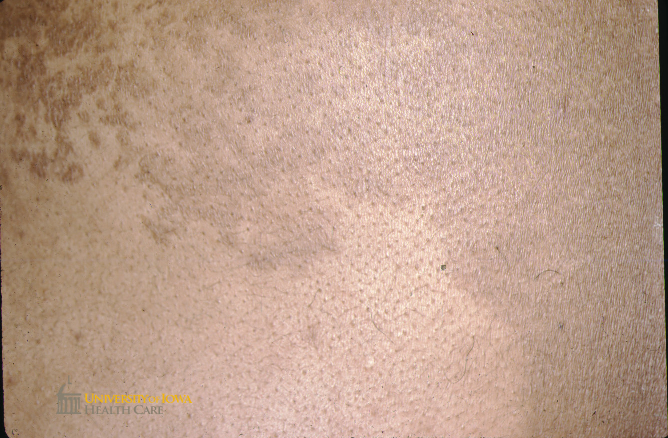
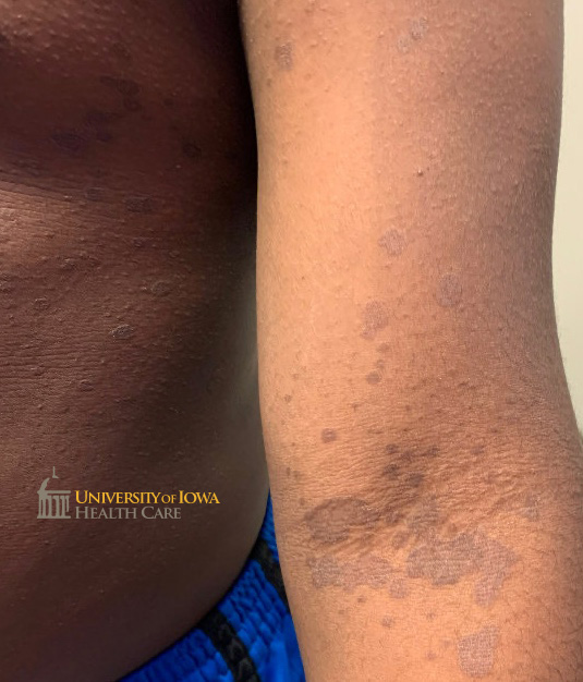
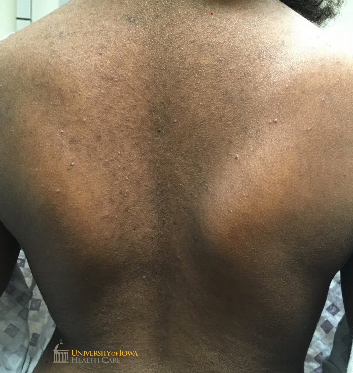
University of Iowa Health Care

Department of Dermatology

Tinea corporis

Fig 1. Hypopigmented slightly scaly thin confluent plaques on the posterior neck, ear, and back. (Click images for higher resolution)

Hypopigmented slightly scaly thin confluent plaques on the posterior neck, ear, and back. (click images for higher resolution).


Fig 2. Hypopigmented slightly scaly thin confluent plaques on the posterior neck, ear, and back. (Click images for higher resolution)

Hypopigmented slightly scaly thin confluent plaques on the posterior neck, ear, and back. (click images for higher resolution).


Fig 3. Hyperpigmented slightly scaly papules coalescing into a plaque on the abdomen. (Click images for higher resolution)

Hyperpigmented slightly scaly papules coalescing into a plaque on the abdomen. (click images for higher resolution).


Fig 4.Gray-brown scaly annular plaques and papules on the trunk and extremity. (Click images for higher resolution)

Gray-brown scaly annular plaques and papules on the trunk and extremity. (click images for higher resolution).

Fig 5.Widespread hyperpigmented scaly papules and the back. (Click images for higher resolution)

Widespread hyperpigmented scaly papules and the back. (click images for higher resolution).

last updated: 08/22/2021