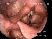
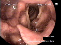
History 73 year old lady with 4-week history of hoarseness. History of smoking and drinking, but none recent. Some swelling of her ankles in the same time-frame as her vocal problems. No other symptoms referable to her head-neck region. Initial examination The patient presents with a moderately dysphonic voice. Her voice is judged overall to be moderately dysphonic, with regular pitch breaks and hoarseness, but retaining a normal pitch range (26 semitones). These characteristics are heard in the passage below.
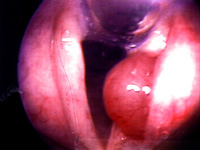
Stroboscopic endoscopic examination shows that neither vocal fold seems to be at all affected by the lesion, and that the visible vibrational interruption is merely from the physical size of the cyst. Some slight damage to the contralateral vocal fold (slight erythma opposite the site of the cyst) may be due to traumatic collision with the cyst. Her vocal cords seem to move normally around the cyst.
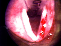
Operation and result Under general anaesthesia, the cyst on the false folds was removed. The lesion was grasped and removed by microscissors. The lesion was sent for pathological analysis and was found to be a laryngeal cyst with papillary oncocytic changes. At her two-month followup, there was no evidence of cyst recurrence, and the patient's voice had returned to normal.
|