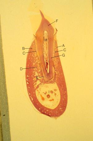
- K-slide 1:
- A. Cortical plate [Buccal]
- B. Cortical plate [Lingual]
- C. Alveolar bone proper
- D. Spongiosa
- E. Periodontal ligament
- F. Coronal dentin
- G. Radicular dentin
- H. Pulp (coronal and radicular)
- I. Inferior alveolar canal
- Note how the PDL narrows near the middle to lower one-third of the root. This indicates the area of the tooth's fulcrum through which tipping (tilting) takes place.
- next
return to index - A. Cortical plate [Buccal]