previous / next
return to index
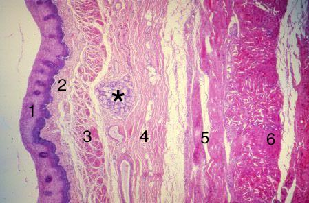
VII-35, Slide 53, Esophagus (H&E). This is a low power view through the wall of the esophagus. Note the following layers: nonkeratinized stratified squamous epithelium (1), lamina propria (2), muscularis mucosae (3), submucosa (4) with esophageal glands (*), inner circular layer (5) and outer longitudinal layer (6) of the muscularis externae. The muscularis externae contains both smooth and skeletal muscle.