previous / next
return to index
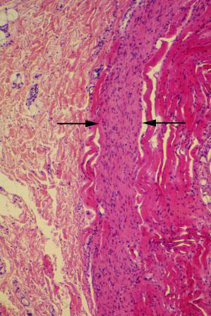
V-67 Esophagus (middle 1/3). Medium power view of submucosa (left) and muscularis externa (inner circular layer, right). Observe collagen fibers in submucosa. Muscularis externa of the middle 1/3 of esophagus consists of both skeletal and smooth muscles; a good place to compare the two muscle types. Smooth muscle is between the arrows. Skeletal muscle stains more eosinophilic, and smooth muscle more basophilic. This is seen to better advantage at higher power in the next slide.